Ribosomes are essential cellular particles found in all living cells. These tiny but mighty structures are the workhorses of protein synthesis, a fundamental process for life. Think of ribosomes as miniature protein factories, diligently translating genetic code into the proteins necessary for cell function and survival. Without ribosomes, cells could not produce the enzymes, structural components, and signaling molecules they need to operate.
The Discovery of Ribosomes
The story of ribosomes began in the mid-20th century with the pioneering work of cell biologist George E. Palade. In 1955, Palade identified small particles, often associated with the rough endoplasmic reticulum in eukaryotic cells. These particles, initially referred to as microsomes, were later recognized and named ribosomes. Palade’s groundbreaking research, utilizing electron microscopy, revealed the ubiquitous nature of ribosomes and hinted at their crucial role within the cell. His discovery earned him the Nobel Prize in Physiology or Medicine in 1974, solidifying the importance of ribosomes in our understanding of cellular biology.
Ribosome Location: Free and Bound Ribosomes
Ribosomes are not confined to a single location within the cell; they are strategically positioned based on the destination of the proteins they produce. In both prokaryotic and eukaryotic cells, ribosomes can be found freely floating in the cytoplasm. These “free ribosomes” synthesize proteins that are destined to function within the cytoplasm itself.
In eukaryotic cells, ribosomes also attach to the membranes of the endoplasmic reticulum, forming the rough endoplasmic reticulum (RER). These “bound ribosomes” are responsible for producing proteins that are destined for secretion outside the cell, insertion into cell membranes, or delivery to organelles like lysosomes. This strategic distribution ensures that proteins are synthesized and directed to their correct locations within or outside the cell.
Ribosome Structure: Subunits and Composition
Despite their small size (around 20-30 nanometers), ribosomes are complex structures composed of two subunits: a large subunit and a small subunit. Each subunit is made up of ribosomal RNA (rRNA) molecules and ribosomal proteins.
In prokaryotes, ribosomes are composed of about 60% rRNA and 40% protein. Eukaryotic ribosomes have a slightly different composition, with roughly equal amounts of rRNA and protein. The subunits are characterized by their sedimentation rate in a centrifugal field, measured in Svedberg units (S). Eukaryotic ribosomes have a large 60S subunit and a small 40S subunit, while prokaryotic ribosomes have a 50S large subunit and a 30S small subunit. These subunits come together to perform the critical task of protein synthesis.
The Function of Ribosomes: Protein Synthesis Explained
The primary function of ribosomes is protein synthesis, also known as translation. This intricate process involves decoding the genetic information carried by messenger RNA (mRNA) to assemble amino acids into polypeptide chains, which then fold into functional proteins. Let’s break down the key steps:
Decoding the Genetic Message (mRNA)
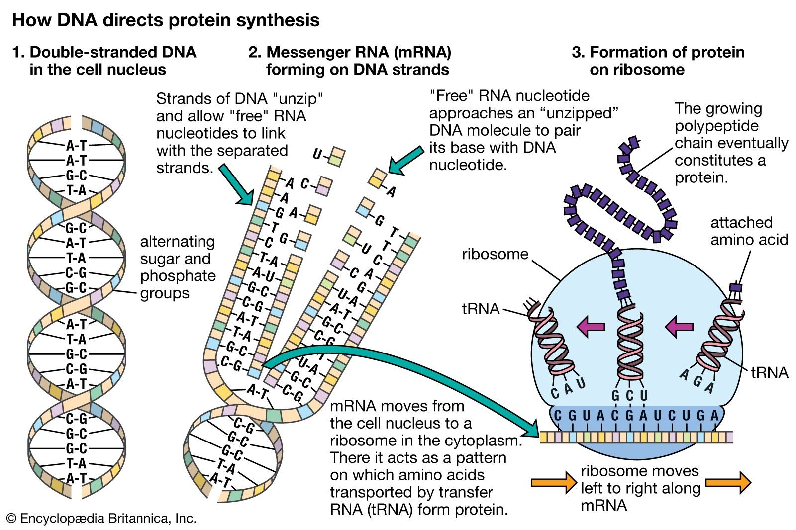
The process begins in the cell nucleus, where DNA serves as the template for creating mRNA. mRNA carries the genetic code, a sequence of nucleotides that dictates the amino acid sequence of a protein. This mRNA molecule travels from the nucleus to the cytoplasm, where it encounters ribosomes. The ribosome binds to the mRNA and moves along it, “reading” the genetic code in triplets of nucleotides called codons.
Assembling Amino Acids (tRNA)
Transfer RNA (tRNA) molecules play a crucial role in bringing the correct amino acids to the ribosome. Each tRNA molecule is attached to a specific amino acid and has a region called an anticodon that can recognize and bind to a complementary codon on the mRNA. As the ribosome moves along the mRNA, tRNA molecules, guided by the codon-anticodon pairing, deliver amino acids in the correct sequence.
Catalyzing Peptide Bonds (rRNA)
Within the ribosome, rRNA plays a catalytic role. Specifically, rRNA catalyzes the formation of peptide bonds between the amino acids brought by tRNA molecules. These peptide bonds link the amino acids together, creating a growing polypeptide chain. As the ribosome continues to move along the mRNA, adding amino acids one by one, the polypeptide chain elongates. Once the ribosome reaches a “stop” codon on the mRNA, protein synthesis is complete. The newly synthesized polypeptide chain is released from the ribosome and folds into its functional three-dimensional protein structure.
 Diagram illustrating the process of protein synthesis, starting with DNA in the cell nucleus and ending with the creation of proteins at ribosomes.
In conclusion, ribosomes are indispensable cellular machines responsible for protein synthesis. Their intricate structure and coordinated function ensure the accurate translation of genetic information into the proteins that drive all cellular processes. Understanding “what ribosomes do” is fundamental to comprehending the very basis of life itself.