In the realm of medicine, the term “lesion” refers to an area of tissue that has been damaged through injury or disease. When we talk about lesions, particularly in the context of neurological conditions like Multiple Sclerosis (MS), we’re focusing on specific areas of harm within the brain or spinal cord. These lesions are a hallmark of MS and understanding what they are, how they form, and their implications is crucial for comprehending the disease itself.
Lesions in Multiple Sclerosis: Plaques of Damage
In Multiple Sclerosis, lesions are frequently referred to as plaques. These plaques are the result of a misguided immune system response. MS is an autoimmune disease where the body’s immune system mistakenly attacks the myelin sheath. Myelin is a protective fatty coating that surrounds nerve fibers in the brain and spinal cord, acting much like insulation around an electrical wire. When the immune system targets myelin, it triggers inflammation and damage. This damage leads to scarring, a process also known as sclerosis. Consequently, these areas of inflammation and scarring are what we identify as lesions or plaques in MS.
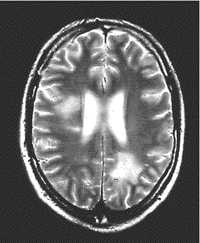 Brain MRI scan
Alt text: MRI scan of a brain showing white spots indicating lesions caused by Multiple Sclerosis.
Detecting Lesions: The Role of MRI Scans in Diagnosis
Magnetic Resonance Imaging, or MRI, is an indispensable tool for detecting and characterizing MS lesions. An MRI scan can not only identify the presence of lesions within the brain and spinal cord but also differentiate between active and inactive lesions. This distinction is vital for understanding the current disease activity.
Active lesions are areas where inflammation is ongoing. To visualize these active lesions on an MRI, a contrast agent containing gadolinium is often used. When injected, gadolinium highlights areas of active inflammation, causing them to appear as bright white patches on the MRI scan. If a lesion “lights up” with gadolinium contrast, it indicates recent inflammatory activity. Conversely, lesions that do not enhance with gadolinium are typically older, inactive lesions, generally more than three months old. An MRI scan performed with gadolinium is often termed an “MRI scan with contrast.”
The number, location, and characteristics of lesions observed on an MRI scan are critical pieces of information for neurologists diagnosing MS. These lesion findings are a significant component of the McDonald Criteria, the internationally recognized diagnostic criteria used to confirm a diagnosis of Multiple Sclerosis.
Are All Lesions Indicative of MS? Understanding Differential Diagnosis
It’s important to recognize that not every lesion detected on an MRI scan is due to Multiple Sclerosis. Lesions are not exclusive to MS and can arise from various other conditions. Migraines and strokes, for instance, can also cause brain lesions. Furthermore, the development of lesions can sometimes be a consequence of the aging process. In some instances, lesions may be incidentally discovered during an MRI scan conducted for an unrelated medical reason, even before any symptoms are experienced. This situation might lead to a diagnosis of Radiologically Isolated Syndrome (RIS). RIS is characterized by the presence of MS-like lesions on MRI in individuals who have not yet experienced clinical symptoms of MS.
While neurologists can sometimes correlate specific symptoms to a particular lesion’s location, this isn’t always straightforward. The size and position of a lesion on an MRI scan don’t always directly correspond to the severity or type of symptoms a person experiences with MS. However, through regular MRI monitoring, neurologists can assess the ongoing activity of MS and gain insights into how the disease might be affecting an individual over time.
Interestingly, lesions are not always permanent. In some cases, the body can repair the damage, and lesions may disappear on subsequent MRI scans. However, persistent lesions can evolve into what are known as ‘black holes’ on MRI. Black holes represent areas where the underlying nerve cells (neurons) have suffered irreversible damage.
Smouldering MS: Chronic Active Lesions and Disease Progression
While some lesions may resolve or remain stable, others exhibit a different behavior. Certain lesions do not heal; instead, they tend to expand slowly over time. When visualized on MRI scans, these growing lesions often display a dark rim of activity around the edges of the damaged area. These are termed chronic active lesions or smouldering lesions. In research settings, they might also be referred to as paramagnetic rim lesions.
Smouldering lesions are increasingly recognized as significant contributors to MS disease progression and the worsening of symptoms over time. The dark rim observed in advanced MRI scans is attributed to microglia. Microglia are immune cells resident in the central nervous system that normally function to clear away damaged cells and debris. However, in MS, it’s believed that the immune system’s attack on nerve cells overactivates microglia within these smouldering lesions. This overactivation leads to chronic inflammation and further damage to nerve cells, contributing to the expansion of the lesion and ongoing neurological decline.
Research has shown a correlation between smouldering lesions and MS progression. Studies have indicated that individuals with four or more smouldering lesions are at a higher risk of developing progressive forms of MS and may experience cognitive and mobility issues at an earlier age compared to those without smouldering lesions. It’s noteworthy that studies have found smouldering lesions in a significant proportion of people with MS, regardless of the specific type of MS they have.
The concept of smouldering MS is shaping current research directions in MS therapeutics. Current disease-modifying drugs (DMDs) primarily target the initial inflammatory attacks on nerves, akin to firefighters tackling the visible flames of a forest fire. However, these treatments may not fully address the underlying, persistent inflammation within smouldering lesions. Newer drugs under development for MS are specifically aimed at targeting this “smouldering burn,” aiming to modulate microglia activity, halt the enlargement of existing lesions, and potentially promote lesion repair.
References
Absinta M, et al. Association of chronic active multiple sclerosis lesions with disability in vivo. JAMA Neurology 2019 Aug 12. Summary (link is external)
Giovannoni, G et al. Smouldering multiple sclerosis: the ‘real MS’ Ther Adv Neurol Disord. 2022; 15: 17562864211066751. Full text (link is external)