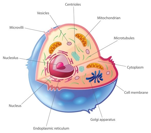
Within the intricate world of a cell, life’s fundamental unit, lie specialized subunits known as organelles. Just as organs perform specific functions within the human body, organelles are the functional units within a cell, each diligently carrying out essential tasks that are vital for cellular survival and activity. These microscopic structures are the workhorses of the cell, orchestrating a symphony of biological processes.
Let’s delve into some key organelles, exploring their structures and the crucial roles they play:
Key Organelles and Their Functions
Nucleus: The Cell’s Control Center
The nucleus stands as the defining feature of eukaryotic cells, distinguishing them from their prokaryotic counterparts. This organelle serves as the cell’s command center, meticulously controlling all cellular activities. Most importantly, it houses the cell’s genetic blueprint – DNA. Within the nucleus, the intricate processes of DNA replication and transcription, crucial steps in gene expression, take place, allowing for precise regulation of genetic information in eukaryotes. While protein synthesis initiation begins in the cytoplasm, the nucleus dictates the process.
Cell Wall: Structural Support and Protection
Enclosing the cells of bacteria, algae, fungi, and plants, the cell wall is a rigid, protective outer layer. This structure is paramount in defining cell shape and providing robust tensile strength, offering critical structural support. Furthermore, the cell wall acts as a shield, protecting the cell from internal osmotic pressure changes and external damage.
Interestingly, bacterial cell walls exhibit diversity, classifying bacteria as gram-positive or gram-negative. Both types rely on peptidoglycan in their cell walls, but gram-negative bacteria possess a more complex structure, featuring a cell wall sandwiched between a plasma membrane and an outer permeable membrane. Gram-positive bacteria, in contrast, have a simpler structure with a single plasma membrane encased by a thicker peptidoglycan cell wall.
Eukaryotic cell walls, found in fungi, algae, and plants, are primarily composed of polysaccharides. For example, fungal cell walls are rich in chitin, while plant and algal cell walls are predominantly built from cellulose microfibrils, highlighting the varied compositions tailored to each organism’s needs.
Centrioles and Centrosomes: Cell Division Organizers
Centrioles are predominantly found in animal cells and, less commonly, in some lower plant cells. Each centriole is a fascinating assembly of nine short microtubule triplets, intricately arranged into a cylindrical shape. Typically, two centrioles are positioned together, forming a centrosome.
Centrosomes are pivotal in cell division (mitosis). They act as the primary microtubule organizing center, essential for constructing the mitotic spindle, which is crucial for chromosome separation during cell division.
Chloroplasts: Powerhouses of Plant Cells
Chloroplasts are double-membraned organelles, uniquely found in plant cells and algae. They bear functional similarities to mitochondria in animal cells, notably in energy conversion. The chloroplast’s outer membrane is freely permeable to small molecules due to porins, whereas the inner membrane regulates molecular traffic via specific transporters.
Chloroplasts possess a unique third membrane system – the thylakoid membrane, essential for the light-dependent reactions of photosynthesis. This membrane is the site of the electron transport chain, crucial for generating ATP, the cell’s energy currency.
The primary function of chloroplasts is photosynthesis. They are responsible for the chemiosmotic reactions that convert carbon dioxide and water into carbohydrates, along with other vital macromolecules like amino acids and fatty acids.
Cilia and Flagella: Movement and Motility
Cilia and flagella are dynamic cell protrusions specialized for movement. Flagella, typically longer and fewer in number, propel entire cells through their environment. Cilia, shorter and more numerous, often work to move substances across the cell surface.
Both cilia and flagella share a common structural motif: a cylindrical arrangement of nine doublet microtubules (composed of one complete and one partial microtubule). Additionally, they feature two central microtubules, completing the “9+2” arrangement characteristic of these motility organelles.
Motion in cilia and flagella is driven by ATP and the sliding of microtubules against each other. This movement is powered by dynein, a motor protein that forms cross-bridges extending from the complete microtubule of one doublet to the partial microtubule of an adjacent doublet, facilitating the sliding mechanism.
Endoplasmic Reticulum (ER): Protein and Lipid Synthesis
The endoplasmic reticulum (ER) is a vast network of membranes within eukaryotic cells, existing in two primary forms: smooth ER and rough ER. The defining structural difference lies in the presence of ribosomes on the outer surface of the rough ER, giving it a ‘rough’ appearance under a microscope.
Rough ER is critically involved in protein synthesis, particularly for proteins destined for secretion or insertion into membranes. Ribosomes attached to the rough ER translate mRNA into proteins. In contrast, smooth ER lacks ribosomes and plays a key role in lipid synthesis, as well as detoxification processes.
Golgi Apparatus: Processing and Packaging Center
The Golgi apparatus, or Golgi complex, is another vital organelle in eukaryotic cells, consisting of flattened, membrane-bound sacs called cisternae, arranged in stacks. It works in close coordination with the ER. The Golgi receives newly synthesized macromolecules, primarily proteins and lipids, from the ER.
Within the Golgi, these molecules undergo further processing, modification, and sorting. The Golgi acts as the cell’s packaging and distribution center, modifying, packaging, and directing proteins and lipids to their final destinations within or outside the cell via vesicles.
Lysosomes: Cellular Recycling and Waste Disposal
Lysosomes are membrane-bound organelles serving as the primary catabolic centers in eukaryotic cells. They are filled with a diverse array of hydrolytic enzymes capable of breaking down all major classes of macromolecules, including polysaccharides, proteins, lipids, and nucleic acids. These enzymes operate optimally at an acidic pH, which is maintained within the lysosome by an ATP-driven proton pump that actively transports protons from the cytoplasm into the lysosome.
Lysosomes are involved in numerous cellular degradation pathways, including phagocytosis (engulfing large particles), endocytosis (internalizing extracellular material), and autophagy (degrading cellular components), allowing for the breakdown and recycling of both external and internal materials.
Mitochondria: Energy Generators
Mitochondria, often referred to as the “powerhouses of the cell,” are double-membraned organelles essential for energy generation in eukaryotic cells.
The inner mitochondrial membrane is highly convoluted, forming cristae, which significantly increase the surface area for ATP production. This inner membrane is impermeable to most small ions and molecules, maintaining the proton gradient crucial for chemiosmosis and ATP synthesis.
Conversely, the outer mitochondrial membrane is permeable to small molecules due to the presence of porins. Beyond energy production, mitochondria are also involved in other critical cellular processes, such as the synthesis of certain steroids, regulation of calcium ion concentration within the cell, and programmed cell death (apoptosis).
Peroxisomes: Detoxification and Metabolism
Peroxisomes are membrane-bound organelles containing a variety of enzymes that catalyze diverse biochemical pathways. They are particularly noted for their role in oxidation reactions, including the breakdown of hydrogen peroxide (a toxic byproduct of metabolism, which is broken down by catalase within peroxisomes), as well as the metabolism of amino acids, uric acid, and fatty acids.
Ribosomes: Protein Synthesis Machines
Ribosomes are not membrane-bound organelles, but rather macromolecular machines essential for protein synthesis. Each ribosome is composed of two subunits, a small and a large subunit, both made up of ribosomal RNA (rRNA) molecules and proteins. Ribosomes can be found freely floating in the cytoplasm or bound to the rough endoplasmic reticulum’s outer membrane.
Regardless of their location, ribosomes perform the critical function of translating messenger RNA (mRNA) into proteins. They act as the platform where amino acids are linked together in the sequence specified by the mRNA, effectively synthesizing proteins from their constituent amino acid building blocks.
Vacuoles: Storage and Turgor Pressure
Vacuoles are large, membrane-bound, fluid-filled sacs predominantly found in plant and fungal cells. They serve diverse roles, including molecular degradation, storage of nutrients and waste products, detoxification, and maintaining turgor pressure within the cell.
In plant cells, the central vacuole plays a crucial role in maintaining cell turgidity, exerting pressure against the cell wall to provide structural support and rigidity to the plant. They are also involved in storing water, ions, and various other molecules.
Conclusion
Organelles are indispensable components of cells, acting as miniature organs that execute a wide array of functions essential for life. From energy production and waste disposal to protein synthesis and cellular communication, each organelle contributes to the overall harmony and functionality of the cell. Understanding organelles is fundamental to grasping the complexities of cell biology and life itself.
References
- http://biology.about.com/od/cellanatomy/ss/organelles.htm
- https://en.wikipedia.org/wiki/Organelle
- http://www.ivyroses.com/Biology/Organelles/Organelle-Functions.php
- http://www.ncbi.nlm.nih.gov/books/NBK9874/
- http://www.ncbi.nlm.nih.gov/pmc/articles/PMC4113101/
- http://www.ncbi.nlm.nih.gov/books/NBK26819/
- https://www.ncbi.nlm.nih.gov/
- http://www.ncbi.nlm.nih.gov/books/NBK26894/
- https://www.ncbi.nlm.nih.gov/