Anatomy, a cornerstone of the biological sciences, delves into the identification and description of the body structures of living organisms. In its broadest sense, it encompasses everything from the visible organs to the microscopic cells that make up life. Let’s explore What Is Anatomical by examining its different branches and historical evolution.
Gross Anatomy: A Macroscopic View
Gross anatomy, also known as macroscopic anatomy, focuses on the study of major body structures that can be observed without the aid of magnification. It traditionally involves dissection and observation to understand the organization and relationships of different body parts. While often associated with the human body, gross anatomy also extends to the study of animal structures.
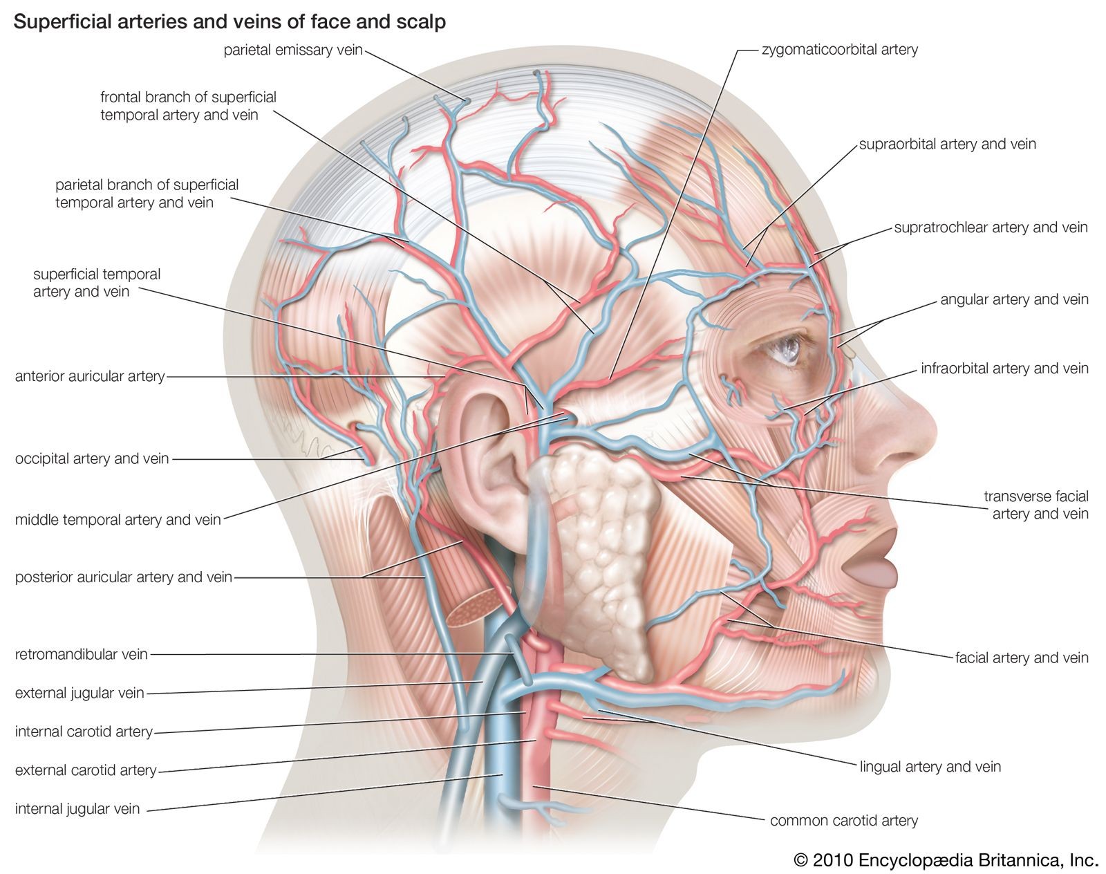 Superficial arteries and veins of the face and scalp, showcasing the intricate network of vessels visible through gross anatomical study
A Historical Perspective
The study of gross anatomy has evolved significantly throughout history. Ancient civilizations, often restricted by cultural and religious beliefs, had limited opportunities for dissection. However, knowledge of the body was gradually acquired through treating injuries, assisting in childbirth, and setting broken bones.
The Alexandrian medical school, particularly under Herophilus (flourished 300 BCE), marked a turning point. Herophilus conducted human dissections, laying a factual foundation for anatomical study. His work was furthered by Erasistratus, sometimes considered the founder of physiology.
In the 2nd century CE, Galen, a Greek physician, compiled and organized the discoveries of earlier Greek anatomists, adding his own physiological concepts and experimental medicine findings. Galen’s writings became the authoritative source for anatomy and medicine in Europe for centuries.
During the Middle Ages, European medicine relied heavily on Galen’s works due to restrictions on dissection. However, the Renaissance saw a renewed interest in direct observation. Leonardo da Vinci’s anatomical drawings paved the way for Andreas Vesalius, whose “De humani corporis fabrica libri septem” (1543) revolutionized the field. Vesalius’s comprehensive and illustrated textbook of anatomy, combined with his emphasis on empirical verification, challenged Galen’s authority and established anatomy as a science based on observation.
Advancements Post-Vesalius
Following Vesalius, anatomists in Padua and beyond made significant strides. They studied digestive glands, urinary systems, and reproductive systems in detail. Hieronymus Fabricius, Gabriello Fallopius, and Bartolomeo Eustachio were among the leading Italian anatomists who contributed to both anatomy and physiology. William Harvey’s discovery of blood circulation, for example, relied on Fabricius’s descriptions of venous valves.
Microscopic Anatomy: Unveiling the Cellular World
Microscopic anatomy, or histology, delves into the study of structures too small to be seen with the naked eye. It involves the use of microscopes to examine cells, tissues, and their organization. This branch of anatomy provides a deeper understanding of the body’s building blocks and their functions.
The Rise of Microscopy
The development of magnifying glasses and compound microscopes in the 17th century was crucial for microscopic anatomy. Marcello Malpighi discovered capillaries, Robert Hooke observed cells, and Antonie van Leeuwenhoek studied muscle fibers and spermatozoa. These discoveries shifted the focus from macroscopic structures to the microscopic realm.
Cellular and Tissue Studies
The 18th and 19th centuries saw systematic advancements in microscopy. Improved microscopes with achromatic lenses enhanced resolving power. Matthias Jakob Schleiden and Theodor Schwann’s cell theory (1838–39) established the cell as the fundamental unit of life.
Microtomes, which allow for the creation of thin tissue sections, and synthetic dyes for staining tissues became standard tools. These advancements led to the rise of cytology (the study of cells) and histology (the study of tissues).
Modern Advancements
In the 20th century, the electron microscope enabled anatomists to study subcellular structures in detail. X-ray diffraction contributed to molecular anatomy, providing insights into the structures of molecules within living organisms.
Anatomical Nomenclature: Standardizing the Language
Anatomical nomenclature involves the standardized naming of body parts and structures. Scientific names are typically in Latin, ensuring clarity and consistency in communication.
The Need for Standardization
Over time, the increasing number of anatomical discoveries led to a proliferation of names, causing confusion. By the late 19th century, the need for standardization became evident.
Efforts Towards Uniformity
The German Anatomical Society initiated efforts to standardize nomenclature, resulting in the Basle Nomina Anatomica (1895). This list reduced the number of terms from 50,000 to 5,528. The Paris Nomina Anatomica (1955) further revised and expanded the list.
The Terminologia Anatomica (1998) is the current international standard for human anatomical nomenclature. It recognizes approximately 7,500 terms describing macroscopic structures and is maintained by the International Federation of Associations of Anatomists.
Comparative Anatomy: Understanding Evolutionary Adaptations
Comparative anatomy examines the similarities and differences in the anatomy of different species. It helps us understand how structures have adapted and evolved over time. By comparing anatomical features across species, we can gain insights into evolutionary relationships and the functional significance of different structures.
In conclusion, “what is anatomical” is a broad field encompassing the study of body structures at all levels of organization. From gross anatomy to microscopic anatomy and comparative anatomy, each branch provides valuable insights into the complexity and diversity of life. The standardization of anatomical nomenclature ensures clear communication and facilitates further advancements in this dynamic field.