Cartilage is a vital structural material in the human body, often described as a connective tissue. It’s firm yet significantly more pliable and softer than bone, providing essential support and flexibility throughout the skeletal system and beyond. Understanding What Is Cartilage and its diverse roles is crucial for grasping fundamental aspects of anatomy and physiology.
Composition and Characteristics of Cartilage
Cartilage is not simply a uniform substance; it’s a complex tissue composed of specialized cells known as chondrocytes. These chondrocytes are responsible for producing a substantial extracellular matrix, the framework that gives cartilage its unique properties. This matrix is rich in:
- Collagen fibers: Providing tensile strength and structure.
- Proteoglycans: Complex molecules that attract water, contributing to cartilage’s resilience and ability to withstand compression.
- Elastin fibers: Adding elasticity and flexibility in certain types of cartilage.
A defining characteristic of cartilage is its avascular nature – it lacks blood vessels. This absence of direct blood supply means chondrocytes rely on diffusion of nutrients from the perichondrium, a dense connective tissue layer that surrounds most cartilage, or from synovial fluid in joints. This diffusion-based nourishment system also explains why cartilage has a slower growth and repair rate compared to tissues with rich blood supplies.
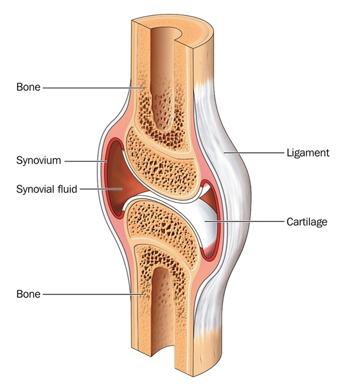 Cross section through a typical synovial joint, showing the bone, synovial membrane, synovial fluid, cartilage and ligament – Image Credit: Blamb / Shutterstock
Diagram illustrating a cross-section of a synovial joint, highlighting cartilage, bone, synovial membrane, synovial fluid, and ligament.
Where is Cartilage Located in the Body?
Cartilage is strategically located in numerous areas throughout the body, performing diverse functions. Key locations include:
- Joints between bones: At articulating surfaces like elbows, knees, ankles, and hips, cartilage acts as a smooth, low-friction surface, facilitating movement and reducing wear and tear between bones.
- Rib Cage: Cartilage connects the ribs to the sternum (breastbone), providing flexibility to the rib cage for breathing.
- Vertebral Discs: Located between the vertebrae of the spine, intervertebral discs made of fibrocartilage act as shock absorbers and allow for spinal flexibility.
- Ears and Nose: Cartilage provides the structural framework for the external ear (auricle) and nose, maintaining their shape and flexibility.
- Respiratory System: Cartilage rings support the trachea (windpipe) and bronchial tubes, keeping airways open for breathing.
Types of Cartilage: Hyaline, Elastic, and Fibrocartilage
Not all cartilage is the same. There are three main types of cartilage, each with distinct properties and functions:
Hyaline Cartilage
Hyaline cartilage is the most abundant type in the body. It is characterized by its pearly bluish-white appearance and a glassy, smooth surface. Key features include:
- Low Friction and Wear Resistance: Ideal for joint surfaces, minimizing friction and wear during movement.
- Weight Distribution: Capable of bearing and distributing weight, crucial in joints like knees and hips.
- Limited Regeneration: Hyaline cartilage has a poor capacity for self-repair when damaged.
Hyaline cartilage is found in:
- Articular surfaces of joints
- Costal cartilage of ribs
- Nasal septum
- Larynx and trachea
Elastic Cartilage
Elastic cartilage stands out due to its remarkable flexibility and elasticity. This type of cartilage contains a high concentration of elastin fibers within its matrix, giving it its springy nature.
Elastic cartilage is found in:
- External ear (auricle)
- Epiglottis (the flap that prevents food from entering the trachea during swallowing)
- Larynx (specifically the cuneiform cartilages)
Fibrocartilage
Fibrocartilage is the toughest and most robust type of cartilage. It is characterized by a dense network of collagen fibers arranged in a less organized manner compared to hyaline cartilage. This structure provides exceptional tensile strength and resistance to compression.
Fibrocartilage is found in:
- Intervertebral discs
- Menisci of the knee
- Pubic symphysis (the joint between the left and right pubic bones)
- Temporomandibular joint (TMJ)
Articular Cartilage: Specialized Hyaline Cartilage in Joints
Articular cartilage is a specific type of hyaline cartilage that covers the ends of bones in synovial joints. Its unique structure is further organized into distinct zones, each contributing to its function:
Zones of Articular Cartilage
-
Superficial Tangential Zone: The outermost layer, making up 10-20% of the thickness. It features:
- Smooth surface for joint gliding.
- Highest collagen content, with fibers aligned parallel to the surface for shear resistance.
- Elongated chondrocytes.
-
Middle (Transitional) Zone: The largest zone, comprising 40-60% of the volume. It exhibits:
- Thicker collagen fibrils, loosely arranged and not parallel to the surface.
- Rounded chondrocytes.
-
Deep Zone: Approximately 30% of the cartilage depth. Characterized by:
- Largest diameter collagen fibrils, oriented perpendicular to the articular surface for compressive strength.
- Highest proteoglycan content and lowest water concentration.
- Columnar arrangement of chondrocytes, parallel to collagen fibers.
-
Calcified Zone: The deepest layer, directly adjacent to the subchondral bone. It contains:
- Small cells within a chondroid matrix.
- Scattered apatitic salts, anchoring cartilage to bone.
Understanding what is cartilage is essential for appreciating its widespread and crucial roles in the body, from enabling smooth joint movement to providing structural support to various organs. Its unique composition and types are perfectly adapted to meet the diverse biomechanical demands placed upon it.