The nervous system is an intricate and vital network within the human body, acting as the primary communication system. It’s responsible for coordinating actions and transmitting signals between different parts of the body. Think of it as the body’s command center and communication highway, working tirelessly to keep everything in sync. From the simplest reflex to the most complex thought, the nervous system is at play.
At its core, the nervous system is composed of a complex collection of nerves and specialized cells known as neurons. These components work together to transmit electrical and chemical signals, allowing for rapid communication throughout the organism. This communication is essential for virtually every bodily function, ensuring that all systems, from the cardiovascular and digestive to the immune system, can effectively interact and maintain overall homeostasis.
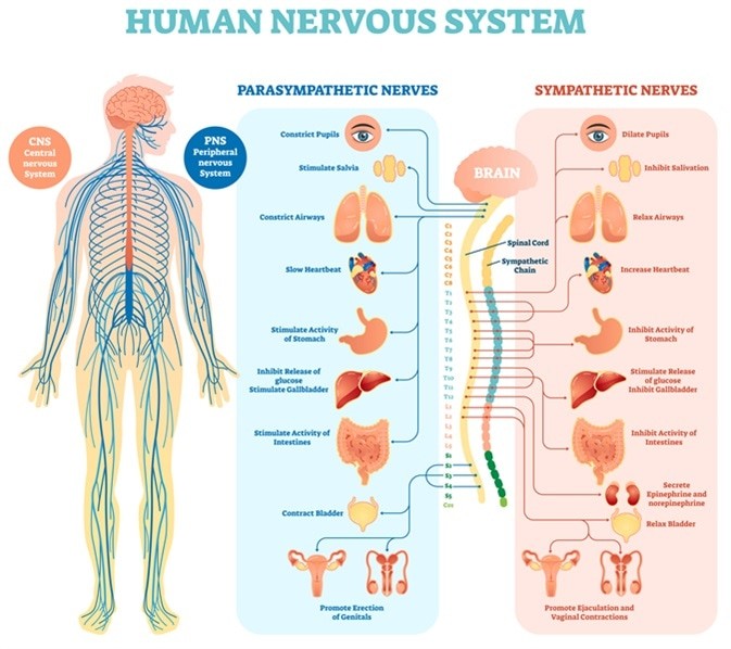
The nervous system is broadly divided into two main parts: the central nervous system (CNS) and the peripheral nervous system (PNS). Understanding this division is key to grasping how this complex system operates. The CNS, the control center, comprises the brain and spinal cord. The PNS, acting as the communication network, extends outwards from the CNS to the rest of the body, encompassing the somatic and autonomic nervous systems.
Image Credit: VectorMine / Shutterstock.com. Depiction of the nervous system’s two main components: the centrally located brain and spinal cord, and the branching peripheral nerves extending throughout the body.
Delving into the Central Nervous System (CNS)
The CNS is the command center of the nervous system, responsible for processing information and coordinating responses. It’s composed of two main structures: the brain and the spinal cord.
The Brain: The Control Hub
The brain, arguably the most complex organ in the human body, is a soft, wrinkled mass weighing, on average, between 1.3 to 1.4 kilograms in adults. Interestingly, around 60% of the brain’s composition is fat, highlighting the importance of lipids in its structure and function. The remaining 40% is a mix of protein, water, carbohydrates, and salts.
This remarkable organ is divided into four principal regions, each with specialized roles:
- Brainstem: Connecting the brain to the spinal cord.
- Cerebrum: The largest part of the brain, responsible for higher-level functions.
- Cerebellum: Located at the back of the brain, crucial for coordination and balance.
- Diencephalon: Situated centrally within the brain, containing the thalamus and hypothalamus.
Together, these areas govern a vast array of functions, including thought, memory, emotions, sensory perception (touch, vision, taste, smell, hearing), motor skills, breathing, hunger, temperature regulation, and countless other processes essential for life.
Within the brain, we find two types of tissue: gray matter and white matter. Gray matter, darker in appearance, is primarily located on the outer surface of the cerebrum and cerebellum (the cortex) and in deeper brain structures called nuclei. It’s largely composed of neuron cell bodies (somas), dendrites, and unmyelinated axons. Gray matter is the brain’s processing center, where information is analyzed and interpreted.
White matter, lighter in color, makes up the inner parts of the brain. It’s mainly composed of myelinated axons – the long, slender projections of neurons that transmit signals. Myelin is a fatty substance that insulates axons and speeds up signal transmission. White matter tracts act as the brain’s communication pathways, connecting different areas of gray matter to each other and to the rest of the nervous system.
Image Credit: ShadeDesign / Shutterstock.com. Illustration showing the structure of a neuron, emphasizing the cell body, branching dendrites for receiving signals, and the axon for transmitting signals.
The Brainstem: Essential Life Functions
The brainstem, a stalk-like structure about an inch long situated at the base of the brain, acts as a crucial bridge connecting the brain to the spinal cord. Despite its small size, it plays a vital role in regulating fundamental life functions. These include:
- Blood pressure regulation
- Breathing control
- Heart rhythm maintenance
- Swallowing coordination
The brainstem is further divided into three main parts:
- Midbrain (Mesencephalon): Involved in eye movements, auditory and visual processing, emotions, and aspects of memory. A key structure within the midbrain is the substantia nigra, rich in dopamine-producing neurons, which is significantly affected in Parkinson’s disease.
- Pons: Serves as a relay station between the cerebrum and cerebellum and is the origin point for four of the twelve cranial nerves. It regulates facial movements, hearing, balance, and breathing.
- Medulla Oblongata: The lowest part of the brainstem, continuous with the spinal cord. It controls vital autonomic functions, including breathing, heart rate, and blood pressure. It also mediates reflex actions like sneezing, vomiting, coughing, and swallowing.
The Cerebrum: Higher Thought and Function
The cerebrum is the largest part of the brain, positioned at the front and comprising the majority of its mass. Its outer layer, the cerebral cortex, is a highly folded sheet of neural tissue, increasing surface area and processing capacity. The cerebrum is divided into two cerebral hemispheres – the right and left – which are connected by a thick band of nerve fibers called the corpus callosum, facilitating communication between them.
While both hemispheres work together, they exhibit some degree of specialization:
- Right Hemisphere: Dominant in spatial awareness, emotions, facial recognition, body posture, and prosody (emotional tone of voice). It also controls the left side of the body.
- Left Hemisphere: Dominant in language processing, logic, reasoning, and processing positive emotions. It controls the right side of the body.
Each hemisphere is further divided into four lobes, named after the skull bones overlying them:
- Frontal Lobe: Located at the front of the brain, responsible for voluntary movement, speech production (Broca’s area), memory, emotions, personality, judgment, planning, organization, and short-term memory.
- Parietal Lobe: Situated behind the frontal lobe and above the occipital lobe. It processes spatial information, allowing us to understand our body’s position in space and relative to objects. It also handles sensory information like pain, touch, temperature, and pressure. Wernicke’s area, crucial for language comprehension, is located in this lobe.
- Temporal Lobe: Located on the sides of the brain, below the parietal lobes. It’s involved in auditory processing, language comprehension (including Wernicke’s area), memory formation (hippocampus is located within), and processing visual and olfactory (smell) information.
- Occipital Lobe: Situated at the back of the brain. It’s primarily responsible for processing visual information, including color, shape, and motion.
The Cerebellum: Balance and Coordination
The cerebellum, meaning “little brain,” is located beneath the temporal and occipital lobes and behind the brainstem. It plays a crucial role in coordinating voluntary motor movements, maintaining balance, and posture. It refines movements, making them smooth and accurate. Emerging research suggests the cerebellum also contributes to cognitive functions like thought, emotion regulation, and social behavior, and may be implicated in conditions like addiction, autism, and schizophrenia.
The Diencephalon: Relay and Regulation
The diencephalon, located deep within the brain, includes two major structures:
- Thalamus: Acts as a sensory relay station, receiving sensory information (except smell) from the body and relaying it to the cerebral cortex for processing.
- Hypothalamus: Plays a vital role in regulating bodily functions like temperature, hunger, thirst, sleep-wake cycles, and hormone release. It communicates with the pituitary gland to control the endocrine system.
The thalamus and hypothalamus, along with the amygdala and hippocampus (located in the temporal lobe), form the limbic system, which is heavily involved in emotion, motivation, memory, and the “fight or flight” response to stress. The hippocampus is particularly crucial for forming new long-term memories, spatial navigation, and learning.
The Spinal Cord: The Information Highway
The spinal cord is a long, cylindrical structure extending downwards from the brainstem through the vertebral column. It serves as the main pathway for communication between the brain and the rest of the body. It’s organized into regions: cervical (neck), thoracic (chest), lumbar (lower back), sacral, and coccygeal.
Thirty-one pairs of spinal nerves branch out from the spinal cord, each pair originating from a specific spinal segment. These nerves carry sensory information from the body to the brain and motor commands from the brain to the muscles and glands.
- Cervical Spinal Cord (8 nerves): Nerves in this region primarily control the head, neck, shoulders, arms, and hands.
- Thoracic Spinal Cord (12 nerves): These nerves control the chest muscles, abdominal muscles, and some back muscles.
- Lumbar Spinal Cord (5 nerves): Nerves in this region control the hips, legs, and feet.
- Sacral Spinal Cord (5 nerves): These nerves control the pelvic organs, bowel, and bladder.
- Coccygeal Spinal Cord (1 nerve): A very small segment at the tail end.
Meninges: Protective Layers
The brain and spinal cord are delicate structures requiring robust protection. This protection is provided by the meninges, three layers of membranes that surround and cushion the CNS. These layers are:
- Dura Mater: The outermost, toughest layer, composed of two sublayers: the periosteal layer (attached to the skull) and the meningeal layer.
- Arachnoid Mater: The middle layer, a web-like membrane made of connective tissue. It lacks blood vessels and nerves. The space between the arachnoid and pia mater, the subarachnoid space, is filled with cerebrospinal fluid (CSF), which further cushions the CNS.
- Pia Mater: The innermost, thinnest layer, tightly adhering to the surface of the brain and spinal cord, following their contours. It is highly vascularized, supplying blood vessels to the CNS tissue.
Neurons: The Building Blocks of the Nervous System
The fundamental unit of the nervous system is the neuron (nerve cell). Neurons are specialized cells designed to transmit information through electrical and chemical signals. They are responsible for all communication within the nervous system.
A typical neuron consists of three main parts:
- Cell Body (Soma): Contains the nucleus and other organelles essential for cell function. It’s the neuron’s control center.
- Dendrites: Branch-like extensions of the cell body that receive signals from other neurons.
- Axon: A long, slender projection extending from the cell body that transmits signals to other neurons, muscles, or glands.
Signals are transmitted electrically down the axon and chemically at synapses, the junctions between neurons. Chemical messengers called neurotransmitters are released at the synapse to transmit signals to the next neuron.
Neurons are broadly classified into three types based on their function:
- Sensory Neurons (Afferent Neurons): Carry sensory information from sensory receptors (e.g., in skin, eyes, ears) to the CNS (brain and spinal cord).
- Motor Neurons (Efferent Neurons): Carry motor commands from the CNS to effector organs (muscles and glands), initiating actions.
- Interneurons (Association Neurons): Located within the CNS, they connect sensory and motor neurons and process information within the CNS. They play a crucial role in complex neural pathways and reflexes.
Many axons are covered in a myelin sheath, an insulating layer made of fat and protein. Myelin is produced by glial cells (Schwann cells in the PNS and oligodendrocytes in the CNS). The myelin sheath speeds up the transmission of electrical signals along the axon. Gaps in the myelin sheath are called Nodes of Ranvier, which also contribute to faster signal transmission through saltatory conduction.
Image Credit: MattLphotography / Shutterstock.com. A visual representation of neurons in a network, highlighting the synapses where neurotransmitters facilitate communication between cells.
Exploring the Peripheral Nervous System (PNS)
The PNS is the network of nerves that lies outside the CNS, connecting the CNS to the rest of the body. It acts as the communication link, relaying information to and from the brain and spinal cord. The PNS is further divided into two main systems: the somatic nervous system and the autonomic nervous system.
The Somatic Nervous System (SNS): Voluntary Control
The somatic nervous system (SNS) controls voluntary movements of skeletal muscles. It’s responsible for our conscious interactions with the external environment. The SNS consists of:
- Sensory nerves: Carry sensory information from the skin, muscles, joints, and sensory organs to the CNS. These sensations include touch, pain, temperature, pressure, and proprioception (body position).
- Motor nerves: Carry motor commands from the CNS to skeletal muscles, causing them to contract and produce movement.
A classic example of SNS function is the reflex response to touching a hot object. Sensory nerves rapidly transmit the pain signal to the spinal cord, which, in turn, triggers motor nerves to command the hand muscles to withdraw, all happening in fractions of a second.
The Autonomic Nervous System (ANS): Involuntary Control
The autonomic nervous system (ANS) regulates involuntary functions, controlling the nerves of internal organs, glands, and smooth muscles. These functions operate largely unconsciously and are essential for maintaining homeostasis. The ANS is further subdivided into:
- Sympathetic Nervous System: Often referred to as the “fight or flight” system. It prepares the body for action in stressful or emergency situations. It increases heart rate, dilates pupils, slows digestion, and releases adrenaline.
- Parasympathetic Nervous System: Known as the “rest and digest” system. It promotes relaxation, conserves energy, and maintains normal body functions. It slows heart rate, stimulates digestion, and constricts pupils.
- Enteric Nervous System: Sometimes considered a third division of the ANS, it’s a network of neurons within the walls of the gastrointestinal tract. It regulates digestion independently of the CNS, although it can be influenced by the sympathetic and parasympathetic systems.
The ANS controls a wide range of vital functions, including heart rate, digestion, breathing, blood pressure, sweating, and sexual arousal, all without conscious effort.
Conclusion: The Indispensable Nervous System
In summary, the nervous system is an incredibly complex and crucial system responsible for coordinating every aspect of bodily function and interaction with the environment. From the central processing of the brain and spinal cord to the far-reaching network of the peripheral nerves, each component plays a vital role in maintaining life, allowing us to perceive, react, think, and feel. Understanding the intricate workings of the nervous system is fundamental to appreciating the complexity and elegance of the human body.