Navigating the intricacies of eye anatomy can be complex, but WHAT.EDU.VN simplifies understanding the “middle structure in the eye,” also known as the uvea or vascular layer. This crucial layer comprises the choroid, ciliary body, and iris, playing a vital role in eye function. Explore this and more on WHAT.EDU.VN for quick, reliable answers and free expert guidance on eye health.
1. Understanding the Eye’s Middle Structure: The Uvea
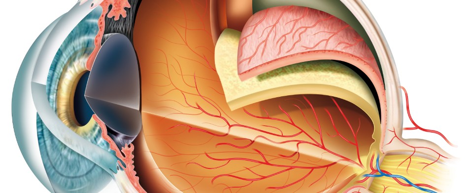
The middle structure in the eye is called the uvea or the vascular layer. The uvea is the eye’s middle layer, located between the sclera (the white outer layer) and the retina (the inner layer that senses light). It consists of three main parts: the choroid, the ciliary body, and the iris.
The uvea is responsible for several critical functions, including:
- Providing blood supply to the retina
- Controlling the amount of light that enters the eye
- Producing the fluid that fills the front of the eye
Understanding the components and functions of the uvea is crucial for maintaining eye health. Let’s explore each part in detail.
2. Components of the Uvea: A Detailed Look
The uvea, or the middle layer of the eye, comprises three integral parts: the choroid, the ciliary body, and the iris. Each component has a specific structure and function, contributing to overall eye health and vision.
2.1. Choroid: Nourishing the Retina
The choroid is the vascular layer located between the sclera and the retina. It is rich in blood vessels that provide essential nutrients and oxygen to the outer layers of the retina. The choroid also contains pigment cells that absorb excess light, preventing internal reflections that could blur vision.
-
Function:
- Nourishes the outer layers of the retina with blood vessels.
- Absorbs excess light to prevent blurring.
 Choroid: Nourishing the Retina
The choroid is crucial for maintaining the health and function of the retina. Conditions affecting the choroid can lead to significant vision problems.
2.2. Ciliary Body: Focusing and Fluid Production
The ciliary body connects the choroid to the iris and has two primary functions: producing aqueous humor and controlling the shape of the lens for accommodation (focusing at different distances).
- Function:
- Produces aqueous humor, which nourishes the cornea and lens.
- Controls the ciliary muscles, which adjust the shape of the lens for focusing.
The ciliary body contains ciliary muscles, which contract and relax to change the shape of the lens, allowing the eye to focus on objects at various distances. This process is known as accommodation. Additionally, the ciliary body produces aqueous humor, a clear fluid that fills the anterior and posterior chambers of the eye, providing nutrients and maintaining intraocular pressure.
2.3. Iris: Controlling Light Entry
The iris is the colored part of the eye, located in front of the lens. It contains muscles that control the size of the pupil, the opening through which light enters the eye.
- Function:
- Regulates the amount of light entering the eye by adjusting the size of the pupil.
- Gives the eye its color.
In bright light, the iris constricts the pupil to reduce the amount of light entering the eye. In dim light, the iris dilates the pupil to allow more light to enter. This mechanism ensures optimal vision under varying light conditions.
3. Importance of the Uvea for Vision
The uvea is essential for maintaining healthy vision. Its components work together to nourish the eye, control light entry, and enable focusing. Understanding its importance can help in recognizing the signs and symptoms of related eye conditions.
3.1. Nourishment and Oxygen Supply
The choroid, as part of the uvea, supplies essential nutrients and oxygen to the retina. Proper nourishment is crucial for the function of photoreceptor cells (rods and cones) in the retina, which are responsible for detecting light and converting it into electrical signals that the brain can interpret.
3.2. Light Regulation
The iris regulates the amount of light that enters the eye, ensuring that the retina is not overwhelmed by excessive light or starved in low-light conditions. This regulation is essential for maintaining clear and comfortable vision.
3.3. Accommodation
The ciliary body controls the shape of the lens, allowing the eye to focus on objects at different distances. This process, known as accommodation, is vital for clear vision at both near and far ranges.
4. Common Conditions Affecting the Uvea
Several conditions can affect the uvea, leading to inflammation, pain, and vision problems. These conditions are collectively known as uveitis.
4.1. Uveitis: Inflammation of the Uvea
Uveitis refers to a group of inflammatory conditions that affect the uvea. It can be caused by infections, autoimmune disorders, injuries, or unknown factors.
- Symptoms of Uveitis:
- Eye pain
- Redness
- Blurred vision
- Light sensitivity (photophobia)
- Dark floating spots in vision (floaters)
Uveitis can affect different parts of the uvea, leading to various types of the condition:
- Anterior Uveitis (Iritis): Affects the iris.
- Intermediate Uveitis: Affects the ciliary body.
- Posterior Uveitis (Choroiditis): Affects the choroid.
- Panuveitis: Affects all parts of the uvea.
4.2. Causes of Uveitis
Uveitis can be caused by various factors, including:
- Infections: Viral, bacterial, or fungal infections.
- Autoimmune Disorders: Conditions like rheumatoid arthritis, lupus, or ankylosing spondylitis.
- Injuries: Trauma to the eye.
- Unknown Factors: In many cases, the cause of uveitis remains unknown (idiopathic).
4.3. Diagnosis and Treatment of Uveitis
Diagnosis of uveitis typically involves a comprehensive eye exam by an ophthalmologist. This may include:
- Visual Acuity Test: Measures how well you can see at various distances.
- Slit-Lamp Examination: Allows the doctor to examine the structures of the eye under high magnification.
- Dilated Eye Exam: Enables a better view of the retina and choroid.
- Blood Tests: To identify underlying infections or autoimmune disorders.
Treatment for uveitis aims to reduce inflammation and relieve symptoms. Common treatments include:
- Corticosteroid Eye Drops: To reduce inflammation in the eye.
- Oral Corticosteroids: For more severe cases or when eye drops are not sufficient.
- Immunosuppressive Medications: To suppress the immune system in cases of autoimmune-related uveitis.
- Antibiotics or Antivirals: If the uveitis is caused by an infection.
4.4. Other Uveal Conditions
Besides uveitis, other conditions can affect the uvea, including:
- Uveal Melanoma: A rare type of cancer that develops in the uvea.
- Uveal Effusion Syndrome: Accumulation of fluid in the uvea, leading to vision problems.
5. Maintaining Uveal Health
Maintaining the health of the uvea is essential for preserving vision and preventing eye problems. Here are some tips to promote uveal health:
5.1. Regular Eye Exams
Regular eye exams are crucial for detecting early signs of uveal conditions. An ophthalmologist can perform a comprehensive eye exam to assess the health of the uvea and identify any potential problems.
5.2. Protect Your Eyes
Protecting your eyes from injuries and infections can help prevent uveitis. Wear protective eyewear during activities that pose a risk of eye injury, such as sports, construction work, or gardening.
5.3. Manage Underlying Conditions
If you have an autoimmune disorder or other underlying condition that can cause uveitis, managing the condition with appropriate medical care is essential. Follow your doctor’s recommendations for treatment and lifestyle modifications.
5.4. Healthy Lifestyle
A healthy lifestyle can contribute to overall eye health, including the uvea. Eat a balanced diet rich in fruits, vegetables, and omega-3 fatty acids. Avoid smoking and limit alcohol consumption, as these can increase the risk of eye problems.
6. Related Structures and Their Roles
To fully appreciate the uvea’s role, it’s helpful to understand the function of related structures in the eye:
6.1. Retina: Light-Sensitive Layer
The retina is the light-sensitive layer at the back of the eye that converts light into electrical signals. These signals are then sent to the brain via the optic nerve, allowing us to see.
- Function:
- Converts light into electrical signals.
- Sends signals to the brain for visual processing.
6.2. Sclera: Protective Outer Layer
The sclera is the white outer layer of the eye that provides protection and support. It is a tough, fibrous tissue that surrounds the eye and attaches to the muscles that control eye movement.
- Function:
- Provides protection and support to the eye.
- Serves as an attachment point for eye muscles.
6.3. Cornea: Clear Front Window
The cornea is the clear, dome-shaped front surface of the eye that helps focus light. It is responsible for about two-thirds of the eye’s focusing power.
- Function:
- Focuses light onto the retina.
- Protects the eye from debris and infection.
6.4. Lens: Fine-Tuning Focus
The lens is a transparent structure located behind the iris that fine-tunes the focus of light onto the retina. It works in conjunction with the cornea to provide clear vision at various distances.
- Function:
- Fine-tunes the focus of light onto the retina.
- Allows the eye to focus on objects at different distances.
7. Latest Research and Studies on Uveal Health
Ongoing research continues to shed light on the complexities of uveal health and the prevention and treatment of related conditions.
7.1. Genetic Factors in Uveitis
Recent studies have identified specific genes that may increase the risk of developing uveitis. Understanding these genetic factors could lead to more targeted treatments and preventive strategies. According to research from the University of California, San Francisco, certain HLA genes are strongly associated with specific types of uveitis.
7.2. New Treatments for Uveitis
Researchers are exploring new treatments for uveitis, including:
- Biologic Therapies: Medications that target specific components of the immune system to reduce inflammation.
- Sustained-Release Implants: Devices that deliver corticosteroids directly to the eye over an extended period.
7.3. Impact of Lifestyle on Uveal Health
Studies have shown that lifestyle factors, such as diet and exercise, can impact uveal health. A diet rich in omega-3 fatty acids and antioxidants may help reduce the risk of uveitis. Regular exercise can also improve overall eye health by promoting healthy blood circulation.
8. FAQ Section on the Middle Structure in the Eye
8.1. What are the first signs of problems in the middle structure of the eye?
Early signs of problems in the uvea, the eye’s middle structure, may include eye pain, redness, blurred vision, increased sensitivity to light (photophobia), and the appearance of dark, floating spots in your vision (floaters). These symptoms can indicate uveitis, an inflammation of the uvea, which requires prompt medical attention to prevent potential vision loss. If you experience any of these symptoms, consult an eye care professional immediately for a thorough examination and appropriate treatment. Regular eye check-ups are crucial for early detection and management of any eye-related issues.
8.2. How can I prevent uveitis?
Preventing uveitis involves several strategies aimed at reducing inflammation and protecting your eyes. This includes managing underlying autoimmune conditions such as rheumatoid arthritis or lupus, as these can trigger uveitis. Protecting your eyes from injury by wearing safety glasses during activities that pose a risk, like sports or construction, is also essential. Maintaining a healthy lifestyle through a balanced diet rich in omega-3 fatty acids and antioxidants may help reduce inflammation. Additionally, practicing good hygiene to prevent infections that could lead to uveitis is important. Regular eye exams can aid in early detection and management.
8.3. Can stress affect the middle structure of the eye?
Yes, stress can indirectly affect the middle structure of the eye. While stress itself may not directly cause uveitis, it can exacerbate underlying conditions that contribute to the inflammation of the uvea. Stress can weaken the immune system, making individuals more susceptible to infections and autoimmune responses, both of which are potential triggers for uveitis. Managing stress through relaxation techniques, regular exercise, and a balanced lifestyle is crucial. Consult a healthcare professional if you experience chronic stress or symptoms of eye problems.
8.4. What is the role of the ciliary body in the middle structure?
The ciliary body is a crucial component of the uvea, the eye’s middle structure. It plays a vital role in producing aqueous humor, a clear fluid that nourishes the cornea and lens. Additionally, the ciliary body contains ciliary muscles, which control the shape of the lens, enabling the eye to focus on objects at varying distances. Proper functioning of the ciliary body is essential for maintaining clear vision and overall eye health. Issues with the ciliary body can lead to conditions such as glaucoma and difficulties with focusing.
8.5. Is uveitis contagious?
Uveitis itself is not contagious. The inflammation of the uvea is typically caused by infections, autoimmune disorders, injuries, or unknown factors, none of which are directly transmitted from person to person. However, if the uveitis is triggered by an infectious agent, such as a virus or bacteria, the underlying infection might be contagious. In such cases, preventive measures to avoid spreading the infection are necessary. Proper diagnosis by an eye care professional is essential to determine the cause of uveitis and implement appropriate management strategies.
8.6. How does diet impact the health of the middle structure of the eye?
Diet plays a significant role in maintaining the health of the uvea. A diet rich in antioxidants, such as vitamins C and E, and omega-3 fatty acids can help reduce inflammation and support overall eye health. Foods like leafy green vegetables, colorful fruits, and fish like salmon and tuna are beneficial. Avoiding processed foods, excessive sugar, and unhealthy fats is also important. Proper hydration supports all bodily functions, including eye health. Nutritional deficiencies can increase the risk of eye problems, so a balanced diet is essential for a healthy uvea.
8.7. What are the long-term effects of uveitis if left untreated?
If left untreated, uveitis can lead to several severe long-term effects on the eye and vision. Chronic inflammation can cause glaucoma, increasing intraocular pressure and damaging the optic nerve. Cataracts, clouding of the lens, can also develop, further impairing vision. Other complications include macular edema, swelling of the macula, and retinal detachment, both of which can result in permanent vision loss. In severe cases, untreated uveitis can lead to blindness. Early diagnosis and treatment are crucial to preventing these complications.
8.8. How is panuveitis different from other forms of uveitis?
Panuveitis is a specific type of uveitis that involves inflammation of all layers of the uvea, including the iris, ciliary body, and choroid. This distinguishes it from other forms of uveitis, such as anterior uveitis (iritis), which affects primarily the iris; intermediate uveitis, which involves the ciliary body; and posterior uveitis (choroiditis), which affects the choroid. Panuveitis often presents with more severe symptoms and can have a broader impact on vision due to the widespread inflammation. Diagnosing panuveitis requires a thorough eye examination to identify the extent of the inflammation.
8.9. Are there any alternative treatments for uveitis?
While conventional medical treatments like corticosteroids and immunosuppressants are the primary methods for managing uveitis, some alternative treatments may help manage symptoms and support overall eye health. These include acupuncture, which some studies suggest can reduce inflammation; herbal remedies like turmeric, known for its anti-inflammatory properties; and dietary changes, such as increasing omega-3 fatty acids. It’s essential to discuss any alternative treatments with your eye care professional before trying them, as they should complement and not replace conventional medical care.
8.10. How often should I get my eyes checked to ensure the health of the middle structure?
The frequency of eye exams to ensure the health of the uvea depends on individual factors, such as age, risk factors, and existing eye conditions. Generally, adults should have a comprehensive eye exam every one to two years. Individuals with a history of uveitis, autoimmune disorders, or other risk factors should have more frequent check-ups as recommended by their eye care professional. Early detection and management of any issues in the middle structure of the eye can help prevent long-term complications. Regular eye exams are a proactive step in maintaining overall eye health.
9. Conclusion: Prioritizing Uveal Health
The uvea, or the middle structure of the eye, plays a vital role in maintaining vision and overall eye health. Understanding its components, functions, and potential problems can help you take proactive steps to protect your eyes. Regular eye exams, a healthy lifestyle, and prompt medical care for any eye symptoms are essential for preserving uveal health.
Have more questions about eye health or any other topic? Visit WHAT.EDU.VN today to ask your questions and receive free, expert answers. Our community of experts is ready to provide you with the information you need, quickly and easily. Don’t hesitate—your answers are just a question away!
Contact Us:
Address: 888 Question City Plaza, Seattle, WA 98101, United States
WhatsApp: +1 (206) 555-7890
Website: WHAT.EDU.VN
Take care of your eyes, and let what.edu.vn help you stay informed and healthy!